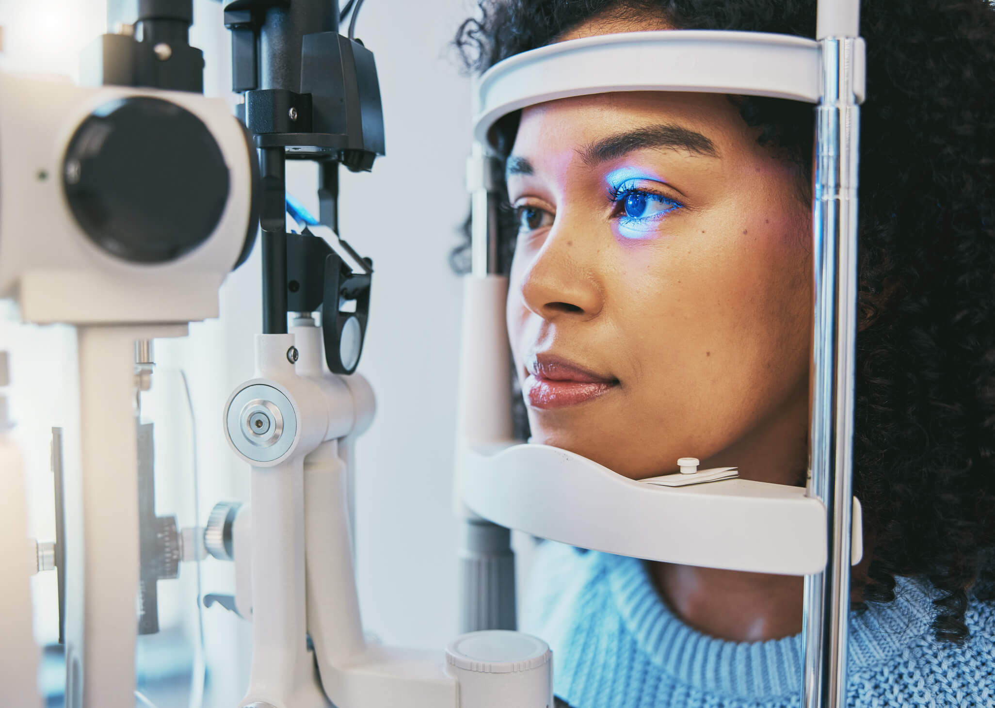
a man getting an eye exam

Can Diabetic Retinopathy Be Reversed? Symptoms, Treatment & Prevention

Diabetic retinopathy cannot be fully reversed, but early-stage cases can be managed to slow or stop progression. Maintaining healthy blood sugar levels and making lifestyle changes can help preserve vision. Advanced stages often require medical intervention to prevent further damage and vision loss.

Many people with diabetes don’t experience noticeable symptoms until the condition has already caused retinal damage. That’s why regular eye exams are essential – they can detect changes in the tiny blood vessels of the retina before significant vision problems occur.

More About Diabetic Retinopathy

Diabetic retinopathy develops when high blood sugar levels damage the blood vessels in the retina, the light-sensitive tissue at the back of the eye. Over time, these blood vessels can leak fluid, become blocked, or lead to the growth of abnormal blood vessels, all of which can cause vision problems.

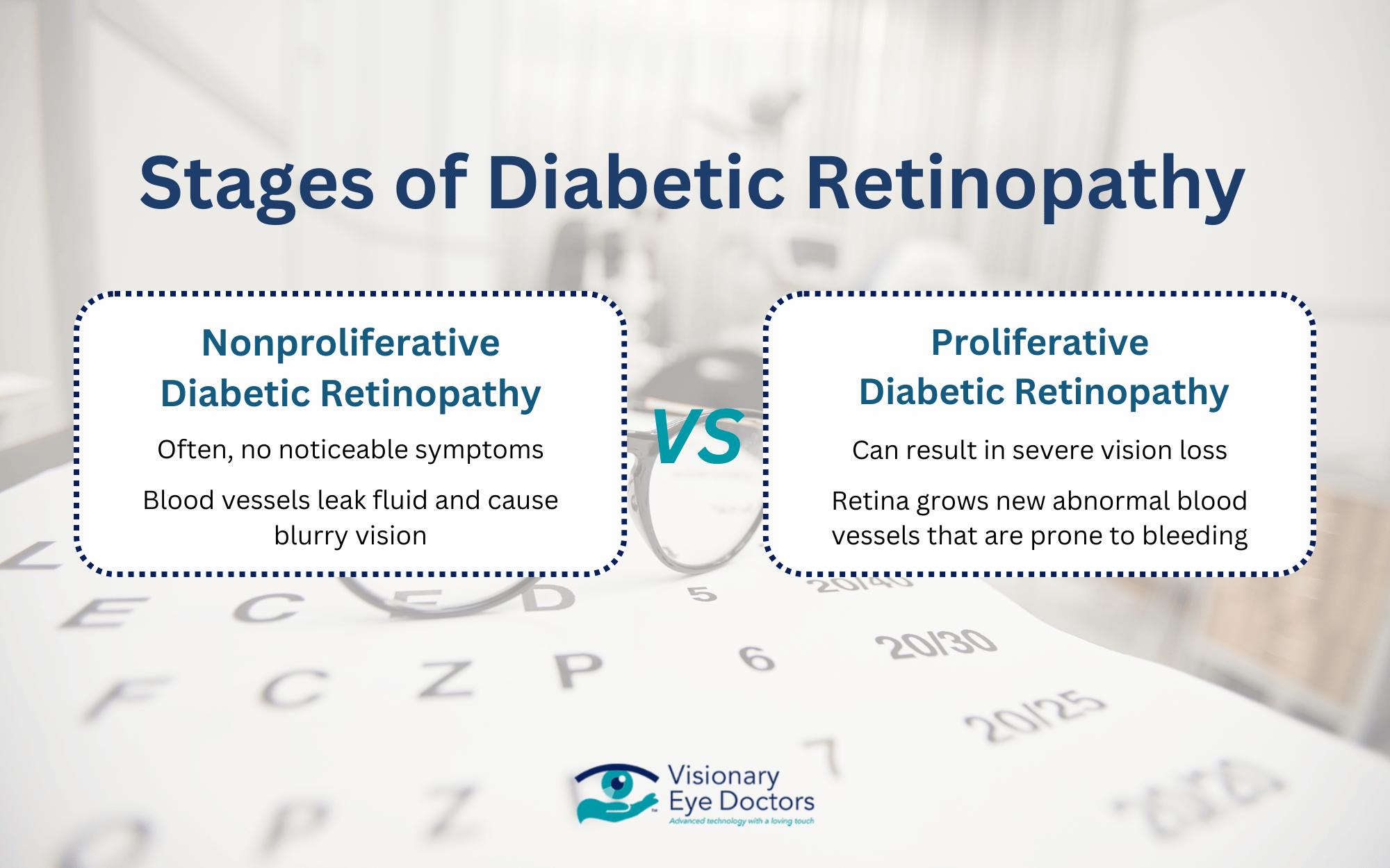
Stages of Diabetic Retinopathy

  • Nonproliferative Diabetic Retinopathy
      • Blood vessels leak fluid, leading to diabetic macular edema that causes blurry vision
      • Many people have no symptoms at this early stage
  • Proliferative Diabetic Retinopathy
    • The retina tries to compensate by growing new blood vessels, but these are fragile and prone to bleeding and scarring
    • Without treatment, this can result in severe vision loss

Managing blood sugar, blood pressure, and cholesterol levels can help slow the progression and protect your vision. Regular eye exams allow for early detection and intervention before significant damage occurs.

Signs and Symptoms

Diabetic retinopathy progresses gradually, with early symptoms including:

  • Blurry vision that fluctuates
  • Floaters  (dark spots or strings in your vision)
  • Difficulty seeing at night or adjusting to dim light
  • Distorted vision, where straight lines appear wavy
  • Dark or empty areas in vision
  • Sudden vision loss (in severe cases)

2 stages of diabetic retinopathy

Is Diabetic Retinopathy Reversible?

Once retinal blood vessels are damaged, the effects are often permanent, particularly in advanced cases where abnormal blood vessels grow or scar tissue forms. However, early intervention can significantly reduce the risk of vision loss and prevent further damage.

Early Stage vs. Advanced Stage

At the early stages of nonproliferative diabetic retinopathy, the focus is on preventing progression through controlling blood sugar, blood pressure, and cholesterol.

When caught early, diabetic retinopathy may not require immediate medical treatment but does require regular eye exams to monitor changes.

In advanced proliferative diabetic retinopathy, blood vessels are abnormal and fragile. At this stage, medical treatment is necessary to prevent permanent vision loss. Treatment methods can include laser therapy, anti-VEGF injections, and vitrectomy surgery.

The Role of Lifestyle Changes in Managing Diabetic Retinopathy

While medical treatments target existing damage, lifestyle changes help prevent worsening symptoms and protect long-term vision health.

  • Blood sugar control reduces the stress of retinal blood vessels
  • Lowering blood pressure and cholesterol prevents further harm to the tiny blood vessels in the eye
  • Regular exercise improves blood flow
  • Antioxidants and omega-3s help limit oxidative stress, which contributes to retinal damage

The Importance of Regular Eye Exams

Because diabetic retinopathy often develops without noticeable symptoms, routine eye exams are essential. Tests like optical coherence tomography (OCT) allow an eye doctor to detect subtle changes in the retina before vision problems appear.

Although this eye condition cannot be reversed, early intervention and consistent management can help preserve vision and prevent severe complications.

In-Depth Treatment Options for Diabetic Retinopathy

While diabetic retinopathy cannot be reversed, several treatments can slow its progression and prevent vision loss. The right approach depends on the severity of the condition and whether diabetic macular edema (DME) or proliferative diabetic retinopathy (PDR) is present.

Non-Surgical Treatments

For early-stage diabetic retinopathy, treatment focuses on stabilizing blood sugar levels, blood pressure, and cholesterol to reduce stress on the retinal blood vessels.

  • Anti-VEGF Injections are injected directly into the eye to block the growth of abnormal blood vessels and help reduce macular edema, potentially stabilizing or improving vision in some patients.
  • Corticosteroid Injections are anti-inflammatory agents that may be used to reduce swelling and inflammation in the retina, especially for those with persistent diabetic macular edema.

Surgical Options

When diabetic retinopathy reaches the advanced stage, surgery may be necessary.

  • Laser Therapy
    • Focal/Grid Laser Treatment targets specific areas of the retina and seals leaking blood vessels to reduce fluid buildup and swelling.
    • Panretinal Photocoagulation targets a wider area of the retina, preventing bleeding and reducing the risk of retinal detachment.
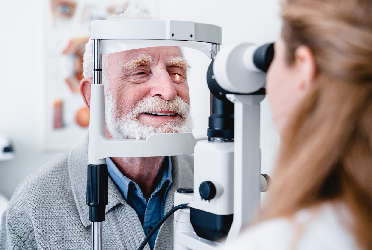
Smiling cheerful elderly patient being checked on eye by female ophthalmic doctor

Protecting Your Vision with Expert Eye Care

Managing diabetic retinopathy requires consistent care and early intervention. While treatments can help slow progression and prevent vision loss, the best way to protect your eyesight is through proactive management and getting regular eye exams at the eye doctor.

Why Ongoing Eye Care Matters

Diabetic retinopathy often develops without noticeable symptoms, making regular eye exams essential. A comprehensive eye exam, including optical coherence tomography (OCT) and dilated retinal imaging, can detect changes in the retina before vision is affected.

How Visionary Eye Doctors Can Help

At Visionary Eye Doctors in the Washington, DC, and Maryland area, our team specializes in diagnosing, monitoring, and treating diabetic retinopathy using advanced technology and personalized care. We provide:

  • Comprehensive eye exams to detect early signs of retinal changes
  • Expert treatment options, including focal and scatter laser treatment
  • Guidance on managing blood sugar, blood pressure, and lifestyle habits to help protect your vision

Take the Next Step in Protecting Your Vision

Don’t wait for noticeable symptoms to appear. Schedule a comprehensive eye exam at Visionary Eye Doctors today to take control of your eye health and prevent any further damage to your vision.

putting contacts in eye

Do I Really Need a Contact Eye Exam Every Year?

Yes, an annual contact lens exam is recommended for most contact lens wearers to protect eye health, maintain an accurate prescription, and detect any underlying vision problems. Even if your vision seems fine, regular exams help prevent issues like dry eye, corneal ulcers, and other eye conditions that may not have obvious symptoms.

Many people believe that once they have a contact lens prescription, they can continue using the same lenses indefinitely. However, vision changes and general wear and tear on the eyes can make old prescriptions ineffective or even unsafe.

The Importance of an Annual Contact Eye Exam

An annual eye exam isn’t just about updating your contact lens prescription – it’s an important step in protecting your eye health.

Unlike glasses, contact lenses sit directly on the eye, making wearers more susceptible to dry eye and irritation. Over time, even a well-fitted contact lens can cause subtle changes to the cornea, which may lead to discomfort if not properly monitored.

Another key reason for regular checkups is that vision changes can occur gradually. Even minor prescription shifts can lead to eye strain, headaches, or blurry vision.

What an Annual Exam Can Detect

Even if you don’t notice any changes in your vision, an annual exam can help detect:

  • Refractive errors that require adjustments for perfect vision
  • Dry eye syndrome
  • Early signs of macular degeneration, diabetic retinopathy, or corneal ulcers
  • Risk factors for vision loss, especially for those with high blood pressure or diabetes

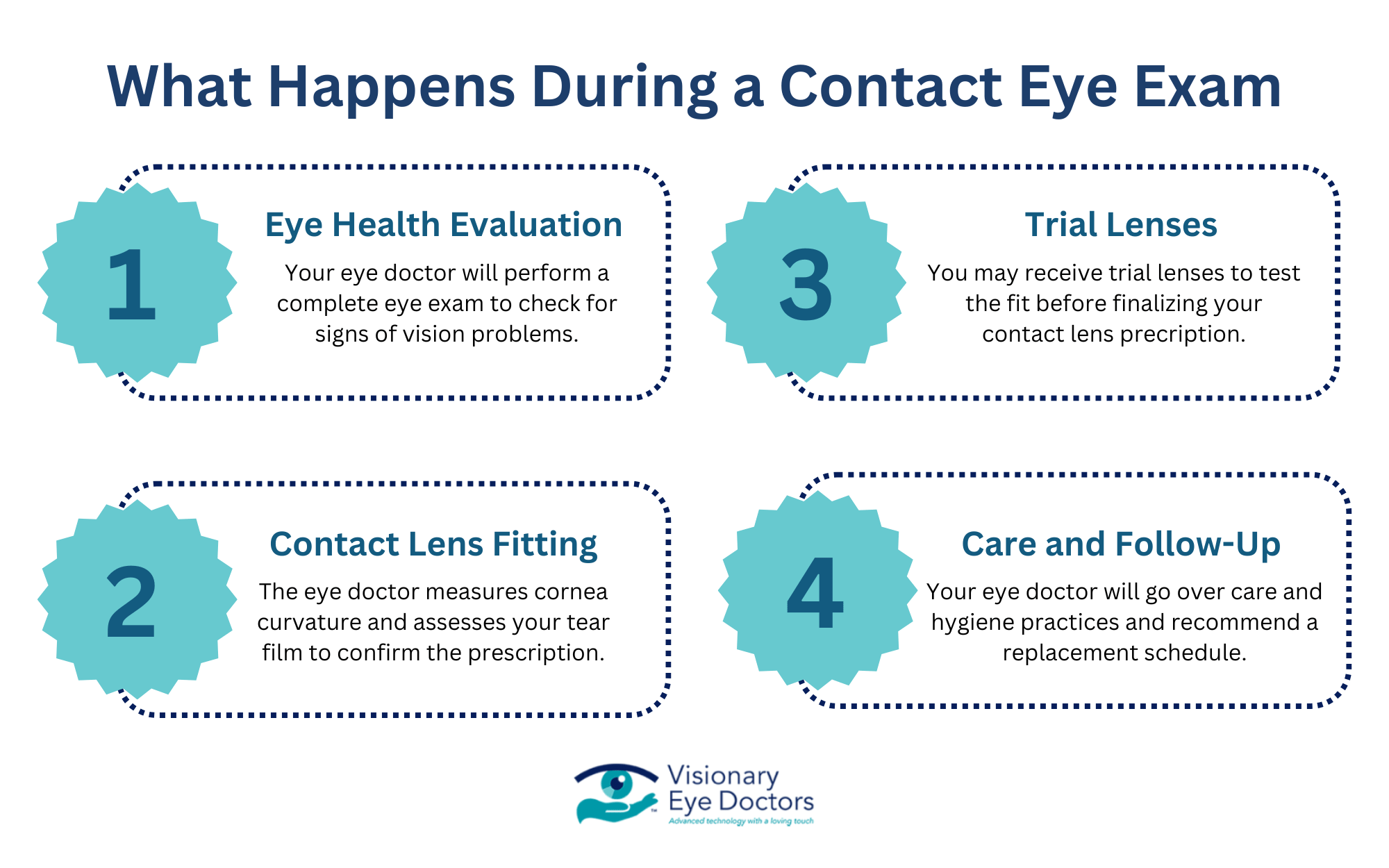
Skipping a routine vision screening increases the risk of wearing an outdated or incorrect lens prescription, which can cause discomfort and vision problems over time. Regular exams provide an opportunity to catch issues early and adjust your vision correction as needed.process of a contact lens exam

What Happens During a Contact Lens Exam?

A contact lens exam is a comprehensive evaluation that ensures your lenses fit properly, your prescription is up to date, and your eye health is protected. Unlike a regular eye exam, a contact lens exam includes additional steps to assess how your lenses interact with your eyes and whether adjustments are needed.

Step 1. Eye Health Evaluation

Before checking your prescription, your eye doctor will perform a complete eye exam to assess your overall eye health. This includes:

  • Checking for vision problems
  • Examining the optic nerve for early stages of glaucoma or macular degeneration
  • Looking for symptoms of dry eye, corneal ulcers, or diabetic retinopathy

Step 2. Contact Lens Fitting

Not all contact lenses are the same, and an improper fit can lead to eye irritation, blurry vision, or discomfort. During this stage, your eye doctor will:

  • Measure the curvature of your cornea to ensure the correct lens fit
  • Assess your tear film to check for dry eye issues
  • Update your contact lens prescription based on your latest vision examination

Even slight changes in your visual acuity can impact how well your lenses perform, which is why keeping your lens prescription current is essential.

Step 3. Trial Lenses and Final Adjustments

If you’re switching to a new type of contact lens, your eye doctor may provide trial lenses to test the fit before finalizing your prescription. They will evaluate:

  • How well the lenses settle on your eyes
  • Whether they provide clear and stable vision correction
  • Any signs of discomfort

Step 4. Care and Follow-Up

To help maintain eye health and prevent infections, your eye doctor will go over:

  • Proper contact lens care and hygiene practices
  • Recommended replacement schedule for your lenses
  • When to schedule your next annual exam or more frequent regular checkups if needed

By the end of the contact lens exam, you’ll have a personalized plan for vision correction that ensures clarity and comfort.

Who Needs More Frequent Contact Lens Exams?

While an annual eye exam is recommended, some individuals may need more frequent checkups to ensure their lenses remain safe and effective.

People at a Higher Risk for Eye Conditions

Signs You Should Schedule an Exam Sooner

Even if you don’t fall into a high-risk group, certain symptoms indicate that you should see an eye doctor sooner:

  • Frequent headaches or blurry vision
  • Redness, irritation, or discomfort while wearing contact lenses
  • Light sensitivity or difficulty seeing at night
  • A history of eye infections

Many vision insurance plans cover annual eye exams, but some policies allow for more frequent checkups for those with higher risk factors. Checking your insurance coverage can help determine whether you qualify for additional regular checkups at little or no extra cost. 

Close up of young woman wearing contact lens with copy space. Young woman holding transparent contact lens on index finger while looking at camera. Close up eye of healthy beautiful girl about to wear contact-lens.

Why Choose Visionary Eye Doctors for Your Next Comprehensive Eye Exam

Your eye health is too important to leave to chance. At Visionary Eye Doctors, we provide comprehensive contact lens exams to protect your vision, comfort, and overall eye health. Whether you need a routine eye exam or adjustments to your prescription, our eye doctors are here to help.

Advanced Technology for Comprehensive Exams

We use the latest diagnostic tools to:

  • Detect early signs of macular degeneration, diabetic retinopathy, and other eye conditions
  • Measure visual acuity and update your prescription for optimal vision correction
  • Evaluate your corneal health to address dry eye concerns

Wide Selection of Contact Lens Options

Not all contact lenses are the same. We offer a variety of lens options, including:

  • Bifocal or multifocal lenses
  • Colored or special-effect contacts
  • Daily disposable lenses
  • Extended wear soft lenses
  • Toric lenses for astigmatism
  • Rigid, gas permeable lenses
  • Scleral lenses
  • Lenses for dry eye
  • Specialty lenses designed specifically for keratoconus

Schedule Your Contact Lens Exam Today

Protect your vision and eye health with a thorough contact lens exam at Visionary Eye Doctors in the Washington, DC and Maryland area. Whether you’re experiencing vision changes, discomfort, or just need an updated prescription, our team is here to provide the best care possible.

Book your appointment today and experience expert eye care tailored to you.

a close up image of a woman's eye

Pinguecula Removal: A Cosmetic Solution for Clearer, Brighter Eyes

If you’ve noticed a yellowish patch on the white part of your eye, you may have a pinguecula. While these growths are typically harmless, they can cause eye irritation, dryness, and a foreign body sensation. For some individuals, a pinguecula’s appearance can be a cosmetic concern.

In such cases, pinguecula surgery is an option for those who want a clearer, more uniform-looking eye. While non-surgical treatments like eye drops can relieve symptoms, surgical excision may be the best choice for a more permanent solution.

What is a Pinguecula and Why Does It Develop?

A pinguecula develops due to changes in the conjunctival tissue, often as a response to long-term exposure to ultraviolet light, wind, dust, and other environmental factors. While older people are more prone to developing pinguecula, it can also appear in younger adults who spend a lot of time outdoors.

normal eye vs pterygium vs pinguecula

Pinguecula vs. Pterygium

While pinguecula and pterygium are often mentioned together, they aren’t the same condition.

  • Pinguecula is a small, raised bump that stays confined to the sclera (the white of the eye). It’s often treated with eye drops or surgical removal for cosmetic reasons.
  • Pterygium (surfer’s eye) is a more advanced growth that extends onto the cornea, potentially affecting vision if it grows large enough. It’s also treated with steroid eye drops or pterygium surgery if vision is affected.

Common Causes of Pinguecula and Pterygium Formation

  • Ultraviolet Radiation: Long-term exposure to ultraviolet rays from the sun is a primary cause of both conditions. UV light can trigger changes in the conjunctival tissue, leading to these growths over time.
  • Environmental Factors: Wind, dust, and dry air can irritate the eyes, increasing the risk of developing a pinguecula or pterygium.
  • Dry Eye and Irritation: Chronic dry eye and frequent eye irritation can contribute to these conditions, as the eye struggles to maintain proper moisture levels.
  • Contact Lens Wearers: Those who wear contact lenses for extended periods may experience increased irritation, which can make a pinguecula or pterygium worse.

Although a pinguecula doesn’t typically affect vision, in severe cases, it may cause discomfort or blurry vision if it becomes inflamed. This condition can lead to redness, irritation, and increased sensitivity.

For many people, a pinguecula is simply a cosmetic concern. Whether it’s due to a noticeable yellowish bump or recurring eye irritation, individuals may want pinguecula treatment to restore the natural appearance and comfort of their eyes.

Treatment Options for Pinguecula: Non-Surgical vs Surgical Removal

Most pinguecula don’t require treatment. However, depending on the severity of symptoms and personal preference, treatment can range from non-surgical approaches to surgical removal.

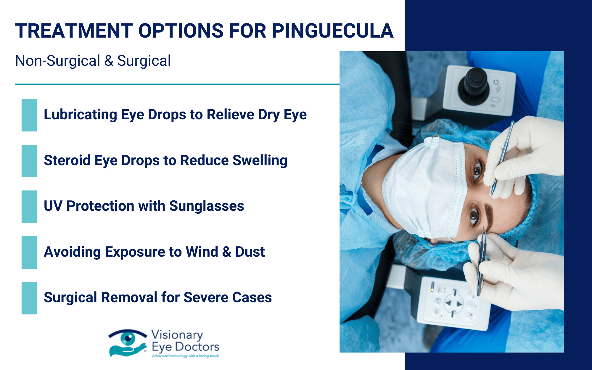
treatment options for pinguecula

Non-Surgical Treatments

In mild to moderate cases, non-surgical treatments can effectively manage symptoms and improve eye comfort:

  • Lubricating Eye Drops: Over-the-counter artificial tears can help relieve dry eye and discomfort.
  • Steroid Eye Drops: If the affected area becomes inflamed or irritated, an eye doctor may prescribe steroid drops to reduce swelling.
  • UV Protection: Wearing UV-blocking sunglasses can help slow progression and prevent further irritation.
  • Avoiding Environmental Triggers: Reducing long-term exposure to wind, dust, and other irritants can help manage symptoms.

Surgical Removal: When Is It Necessary?

For those wanting a long-term solution, surgical excision may be recommended. Proper surgical removal is considered in the following cases:

  • The pinguecula is severely inflamed despite the use of eye drops
  • The pinguecula causes chronic discomfort or blurry vision
  • The patient wants pinguecula surgery for cosmetic reasons to restore a natural appearance to the eye

Pinguecula Surgery: What to Expect

The surgical removal of a pinguecula is a minimally invasive outpatient procedure performed by a corneal specialist or eye surgeon. It involves carefully removing the affected conjunctival tissue while preserving as much healthy tissue as possible.

To improve the cosmetic outcome and reduce the risk of recurrence, advanced techniques may be used:

  • Conjunctival Autograft: A small section of healthy conjunctival tissue is glued over the affected area, promoting smoother healing and improving dry eye.
  • Amniotic Membrane Grafting: An amniotic membrane is used to enhance healing and prevent the pinguecula from returning.
  • Fibrin Glue: To improve patient comfort, fibrin glue is used to secure the graft instead of traditional sutures, leading to a faster recovery time.

Most patients experience minimal downtime after pinguecula surgery. Surgical excision is generally safe, but as with any procedure, there is a small risk of recurrence, redness, or eye irritation. Following post-operative care – using steroid drops – is essential for an optimal healing process.

The Cosmetic Benefits of Pinguecula Removal

Even if a pinguecula isn’t causing significant irritation or redness, some people choose surgical removal to achieve a brighter, healthier-looking eye. Although pingueculas are harmless, they can make the eyes look discolored, red and irritated, and aged or unhealthy.

If your pinguecula is affecting your confidence or quality of life, speaking with an eye doctor can help determine if surgical removal is the best option. For many patients, the combination of advanced surgical techniques and proper aftercare leads to improved appearance with minimal downtime.

a girl getting an eye exam

Why Choose Visionary Eye Doctors for Pinguecula Removal

If you’re considering pinguecula surgery for cosmetic reasons, choosing the right eye doctor is essential. At Visionary Eye Doctors, we combine medical expertise, advanced technology, and patient-centered care to deliver the best possible cosmetic and medical outcomes.

Our highly skilled eye surgeons have extensive experience in pinguecula and pterygium removal, using the latest techniques to ensure:

  1. Precise, safe surgical excision that preserves healthy conjunctival tissue
  2. Minimized scarring and recurrence
  3. Superior cosmetic outcomes, so your eyes look bright, clear, and natural after surgery

A Personalized Approach to Eye Care

We understand that every patient has unique concerns, and that’s why we provide personalized consultations and comprehensive post-surgery care. We discuss all treatment options and provide guidance to help patients prevent future issues.

Schedule a Consultation

If your pinguecula is affecting your appearance or eye comfort, our team at Visionary Eye Doctors is here to help. With our expertise in cosmetic and medical eye care, we offer safe, effective solutions tailored to your needs.

Schedule a consultation today to learn more about how we can help you achieve healthier, brighter-looking eyes!

Resources:

https://www.aoa.org/healthy-eyes/eye-and-vision-conditions/pinguecula 

https://my.clevelandclinic.org/health/diseases/22497-pterygium-surfers-eye

a woman getting glasses

Low Vision Awareness Month: Empowering Your Vision Health

February is Low Vision Awareness Month, a time dedicated to raising awareness about low vision, its causes, and the resources available to help those affected.

Millions of people in the United States experience vision loss due to conditions like age-related macular degeneration (AMD), diabetic retinopathy, glaucoma, and retinitis pigmentosa. While these conditions can significantly impact everyday tasks, regular eye exams and vision rehabilitation can help individuals make the most of their remaining vision.

low vision awareness month graphic

What Exactly is Low Vision?

Low vision is a permanent vision impairment that cannot be fully corrected with regular glasses or contact lenses. Unlike typical vision problems that can be managed with prescription lenses, low vision makes daily activities – such as reading, recognizing faces, and driving – more difficult, even with the best possible corrective eyewear.

Common Causes of Low Vision

Several eye conditions can lead to low vision, including:

  • Age-Related Macular Degeneration: A leading cause of vision loss in older adults, AMD affects central vision, making it difficult to see the fine details.
  • Diabetic Retinopathy: Caused by damage to the blood vessels in the retina, this condition can lead to visual impairment and blind spots.
  • Glaucoma: A group of diseases that damage the optic nerve, often leading to gradual vision loss that may not be noticeable at first.
  • Retinitis Pigmentosa: A genetic disorder that affects night vision and peripheral vision, progressing to significant impairment over time.
  • Cataracts: Gradually clouds the lens of the eye, leading to blurry vision and difficulty seeing at night.
  • Eye Injuries: Caused by a pervious trauma to the eye and retinal detachment.

While these conditions develop for different reasons, early detection through regular eye exams is crucial in slowing their progression and preserving detailed vision for as long as possible.

The Importance of Early Detection and Regular Eye Exams

Detecting vision problems early can make a significant difference in managing low vision and preserving eyesight. Many eye conditions develop gradually and may not show noticeable symptoms until vision loss has already occurred.

How Often Should You Get an Eye Exam?

The American Academy of Ophthalmology and the National Eye Institute recommend eye exams based on an individual’s age and risk factors. The following are guidelines for comprehensive eye exams:

  • Children (before age 18): An initial eye exam at six months, again at age three, and before starting school. After that, exams every 1-2 years help detect vision problems early.
  • Adults under 40: At least every 2 years, unless you have risk factors like diabetes or a family history of vision problems – in which case, annual exams are recommended.
  • Adults 40-60: An exam every year to check for early signs of glaucoma, diabetic retinopathy, and AMD.
  • Adults 60 and older: Annual exams are highly recommended as the risk of vision impairment and blind spots increases with age.

How Regular Eye Exams Can Help

Routine eye exams allow eye care professionals to:

  1. Detect early signs of vision impairment and underlying eye conditions
  2. Monitor risk factors like high blood pressure or diabetes, which can contribute to vision loss
  3. Identify changes in central vision that may indicate macular degeneration
  4. Use tools to check for blind spots or distorted straight lines

For those already experiencing low vision, regular checkups with a low vision specialist can help determine the best medical treatments and preventive measures to improve daily life.

Vision Rehabilitation and Support Resources

For those experiencing low vision, adjusting to daily tasks can be challenging, but vision rehabilitation programs provide the tools, training, and support to help individuals maximize their remaining vision.

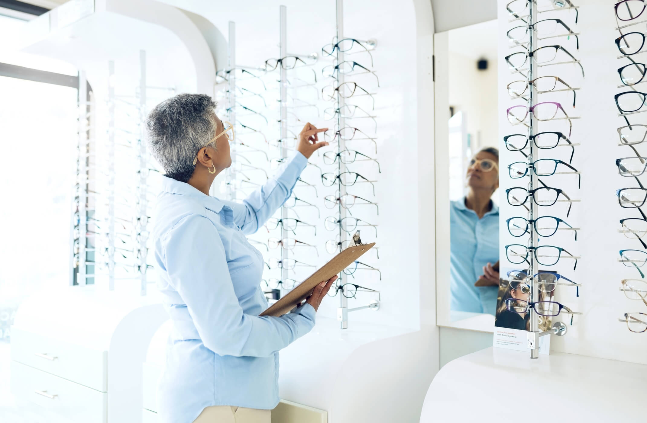
a woman picking out glasses

What is Vision Rehabilitation?

Vision rehabilitation is a personalized approach involving various strategies, low vision aids, and specialized training. A low vision specialist works closely with each patient to develop a plan that meets their specific needs.

Key aspects of vision rehabilitation include:

  • Low Vision Aids: Devices like magnifiers, special lighting, and large-print materials designed for people with vision impairment.
  • Assistive Technology: Screen readers and voice-controlled devices that enhance detailed vision for tasks like reading and writing.
  • Occupational Therapy: Occupational therapists teach strategies like labeling household items, improving contrast in home environments, and using adaptive techniques for cooking and personal care.
  • Mobility Training: Mobility specialists help individuals learn how to safely navigate their surroundings at home and in public.
  • Emotional Support: Adjusting to vision loss can be overwhelming and lead to emotional stress. Connecting with support groups can provide encouragement and practical tips from others facing similar challenges.

Where to Find Help

There are many resources available for those living with low vision:

  • National Eye Institute: Offers information on vision problems, treatments, and resources for people with low vision.
  • Prevent Blindness: A national nonprofit dedicated to eye health education, advocacy, and patient support.
  • American Optometric Association: A professional organization that promotes doctrors of optometry in practice management and development.

By utilizing vision rehabilitation, individuals can maintain their independence while learning skills to make the most of their remaining vision.

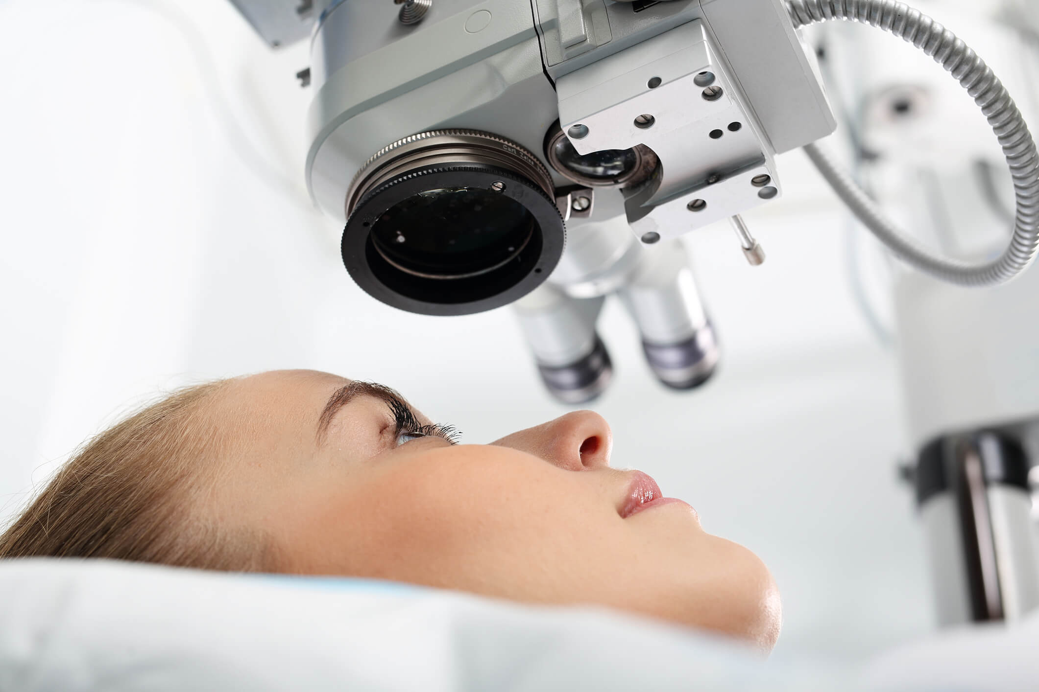
a girl getting an eye exam

Empowering Your Vision Health with Visionary Eye Doctors

Living with low vision presents challenges, but the right care, tools, and support can make all the difference. At Visionary Eye Doctors,  we help individuals with vision impairment maintain their eye health, independence, and quality of life.

Comprehensive Eye Care and Support

Our team of eye doctors offers a full range of services for Glaucoma, Cataracts, Macular Degeneration, Diabetic Retinopathy, Pterygium, Keratoconous, and more:

  • Thorough eye exams that help detect early signs of vision loss or impairment
  • Advanced diagnostic tools and technology to treat various eye conditions
  • Medical treatments and preventative care to help slow progression and preserve vision

Our team works closely with each patient to ensure they have access to the resources and guidance they need to deal with low vision.

Take the Next Step in Protecting Your Vision

Regular eye exams are the first line of defense against vision problems. If you are experiencing symptoms of low vision, don’t wait – early intervention can help preserve your remaining sight and improve your daily life.

Contact Visionary Eye Doctors today to schedule an appointment with one of our eye specialists and explore the best options for protecting your eye health.


Resources:

https://www.aao.org/

https://www.nei.nih.gov/ 

https://www.aoa.org/