The Effects of Macular Degeneration
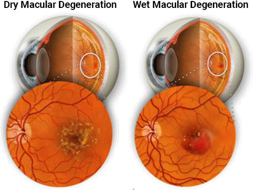
Macular degeneration affects the area of the retina called the macula, the part of the eye responsible for central vision. A person with this condition has difficulty seeing detailed objects such as small print, faces, or street signs. The affected areas of the macula often cause scotomas, or small central areas of vision loss. These areas may cause objects to appear faded, disappear, or look distorted. Straight lines may look wavy. Peripheral vision is not affected and macular degeneration does not cause total blindness.
Possible Causes and Risk Factors
The exact cause of age-related macular degeneration (ARMD) is unknown. What is known by macular degeneration doctors is that there are many risk factors associated with AMD, including:
- Age. As we age our risk of developing AMD increases. The risk of AMD at age 50 is only 2%. That rate jumps to 37% by age 75.
- Family History. One’s risk of developing AMD is 300% higher if an immediate family member has the condition.
- Genetics. A strong association between development of AMD and presence of variant genes including complement factor H (CFH) and complement factor B.
- Smoking. Patients who smoke are 5 to 8x more likely to develop AMD compared to nonsmokers
- Racial predisposition. Caucasians and patients with light-colored skin and eyes are more likely to develop AMD than darker pigmented patients.
- Obesity. High Blood Pressure and High Cholesterol
- UV exposure. It is important to protect your eyes by wearing Sunglasses with 100% UV protection
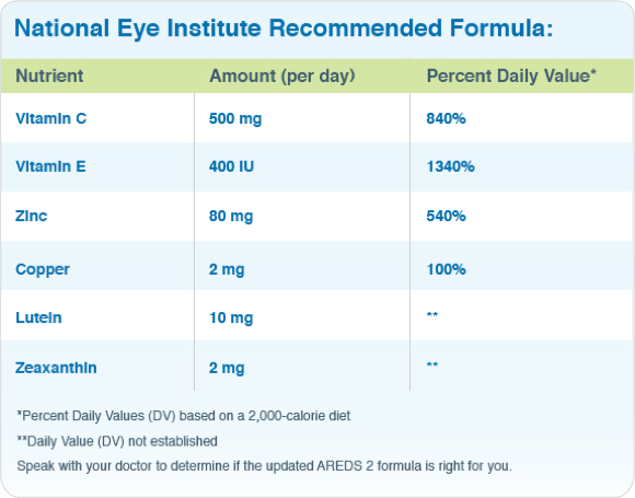
- Diet. Diets high in saturated fats (ie meat, butter, cheese) and low in Vitamin C, E and lutein increase one’s risk factor for AMD
How We Diagnose Macular Degeneration
Routine and yearly dilated ocular examination is important for early management. During your examination, which is done by the best macular degeneration doctors, imaging scans of the retina, called Optical coherence tomography (or OCT for short) is performed, providing detailed imaging of the retina and macula.
In addition, a dye test, called a Fluorescein Angiography maybe performed. A yellow dye (called Fluorescein) is injected into your vein. Travelling through the body and into the eyes where using a special camera, highlights abnormal new vascular growth or leakage helping identify Wet AMD.

